
Health December 15, 2025
Renal Ultrasound and Imaging: How to Evaluate Kidney Obstruction and Size
When your doctor suspects a blockage in your urinary tract or wants to check if your kidneys are the right size, renal ultrasound is usually the first test they order. It’s quick, safe, and doesn’t use radiation-unlike CT scans or X-rays. For many people, especially those with kidney stones, urinary retention, or suspected blockages, this simple scan gives doctors the answers they need without putting you at risk.
What Renal Ultrasound Actually Shows
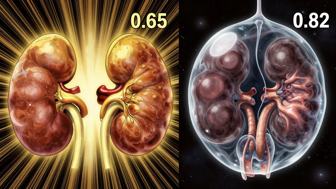
Renal ultrasound uses sound waves to create real-time images of your kidneys, bladder, and the tubes that carry urine (ureters). It doesn’t just show shape-it measures size, thickness, and pressure changes inside the kidney. Normal adult kidneys are about 9 to 13 centimeters long. If one is significantly smaller, it could mean long-term damage. If it’s enlarged, it might be swollen with backed-up urine-a sign of obstruction.
The key thing doctors look for is hydronephrosis. That’s when urine can’t drain properly and starts pooling in the kidney’s collecting system. On ultrasound, this looks like dark, fluid-filled spaces inside the kidney. The more dilation, the worse the blockage. Radiologists grade it using the Society for Fetal Urology system, from mild (slight swelling) to severe (kidney looks like a balloon filled with fluid).
Measuring Kidney Size and Cortical Thickness
Kidney size isn’t just about length. Cortical thickness-the outer layer of functional tissue-is just as important. A healthy cortex is at least 1 centimeter thick. If it’s thinner than that, it often means the kidney has been under pressure for a long time, maybe from chronic obstruction or high blood pressure. In older adults or people with diabetes, thinning cortex can signal early kidney disease even before blood tests show problems.
Doctors measure both kidneys side by side. One kidney might be slightly larger than the other-this is normal. But if one is 2 cm or more shorter than the other, or if the cortex is visibly thinner, it’s a red flag. These measurements help decide if surgery is needed or if the blockage can be managed with medication or monitoring.
How Ultrasound Detects Obstruction
Obstruction usually happens at three places: where the kidney meets the ureter (UPJ), where the ureter crosses the pelvic bone, or near the bladder. Ultrasound doesn’t always see the stone or tumor causing the blockage, but it sees what it does to the kidney.
The real game-changer is Doppler ultrasound. This part of the scan measures blood flow inside the kidney. When urine backs up, pressure builds up, and blood flow slows down. Doctors calculate something called the resistive index (RI)-a number between 0 and 1. A normal RI is below 0.70. If it’s 0.70 or higher, it strongly suggests obstruction. One 2015 study found this method was 86.7% accurate at detecting blockages.
Why does this matter? Because some people have mild hydronephrosis without obstruction. The RI helps tell the difference. A swollen kidney with a low RI might just be a normal variant. A swollen kidney with a high RI? That’s likely blocked-and needs action.
Ultrasound vs. CT and MRI: Why It’s Still First Choice
CT scans are great at finding tiny stones-down to 1 or 2 millimeters. But they expose you to radiation equivalent to 300 chest X-rays. A single CT urogram delivers about 10 millisieverts of radiation. For someone with recurrent kidney stones, that adds up fast. Ultrasound gives you 80% of the info without any radiation.
MRI urography shows amazing detail and can map how urine flows, but it costs 3 to 5 times more than ultrasound-often $1,500 to $2,500-and takes longer. It’s also not great at spotting stones. Nuclear scans measure kidney function but involve radioactive tracers and don’t show anatomy well.
Ultrasound wins on safety, speed, and cost. It costs between $200 and $500 in the U.S., takes 15 to 30 minutes, and doesn’t require fasting or injections. Emergency rooms use it because it’s fast. If you come in with severe flank pain, a bedside ultrasound can rule out obstruction in minutes, cutting down wait times by nearly 45 minutes.
Where Ultrasound Falls Short
It’s not perfect. If you’re overweight-with a BMI over 35-sound waves can’t penetrate well. The images get blurry, and doctors may have to switch to CT or MRI. Bowel gas can also block the view, especially if you’ve eaten recently.
Ultrasound can’t measure how fast urine drains from the kidney. CT scans with special software can track this using contrast dye, but ultrasound can’t. It also doesn’t show small stones well. A 2-millimeter stone might slip past the scan, especially if it’s in the ureter.
And here’s the big catch: it depends on who’s doing the scan. A 2018 study showed up to 20% variation in kidney size measurements between new and experienced sonographers. That’s why training matters. The American Institute of Ultrasound in Medicine requires at least 40 supervised scans before you can perform them independently. Many radiology residents say it takes around 50 exams to feel confident measuring resistive index accurately.
What Happens After the Scan?
If your ultrasound shows mild hydronephrosis and a normal resistive index, your doctor might just watch and wait. You’ll come back in a few weeks for a repeat scan. If the swelling grows or the RI climbs, they’ll look for the cause-maybe a stone, a tumor, or a narrowing in the ureter.
If obstruction is confirmed, treatment depends on the cause. A small stone might pass on its own. A large one may need shockwave therapy or a stent. In children with UPJ obstruction, surgery is often needed to fix the blockage before kidney damage sets in. For older adults, a stent or nephrostomy tube might be placed to drain urine while they recover.
Ultrasound is perfect for follow-up. One urologist told me he monitors post-surgery patients weekly with bedside ultrasound instead of repeating CT scans. No radiation. No cost. Fast results. That’s the real power of this tool.
What’s New in Kidney Ultrasound?
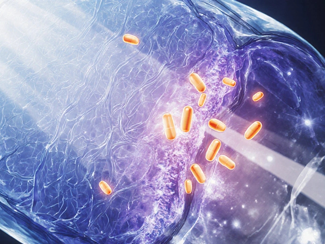
Technology is pushing this field forward. Shear-wave elastography measures how stiff kidney tissue is. When urine backs up, pressure makes the tissue stiffer. This technique can detect early obstruction even before swelling shows up on standard images.
Another breakthrough is super-resolution ultrasound. Researchers are now able to visualize tiny blood vessels inside the kidney that were invisible before. This could help spot early signs of scarring or reduced blood flow-long before kidney function drops.
Artificial intelligence is also entering the picture. Some hospitals are testing AI tools that automatically measure hydronephrosis grade and calculate resistive index. One Mayo Clinic trial showed AI matched expert radiologists 94% of the time. In the future, these tools could help reduce human error and make scans faster.

Who Gets This Test?
Renal ultrasound is standard for:
- People with sudden, severe flank pain (suspected kidney stones)
- Pregnant women with urinary symptoms (radiation is risky for the fetus)
- Children with suspected urinary tract blockages
- Patients with unexplained kidney failure
- Those with a history of kidney stones or surgery
- People with high blood pressure and reduced kidney function
It’s also used in emergency departments, urgent care centers, and even some primary care offices-though only about 35% of primary care clinics have the equipment. Most hospitals have it, and 75% of academic emergency rooms use point-of-care ultrasound for kidney issues.
What to Expect During the Exam
You don’t need to fast. Drink water if you’re being checked for obstruction-it helps fill your bladder and gives better views. You’ll lie on your back or side. The sonographer will put gel on your back or belly and move a handheld probe over your skin. You might feel slight pressure, but no pain.
The whole thing takes 15 to 30 minutes. Results are often available the same day. The report will include:
- Kidney length, width, and thickness
- Cortical thickness
- Anteroposterior diameter of the renal pelvis
- Presence and grade of hydronephrosis
- Resistive index values from both kidneys
If anything looks off, your doctor will explain what it means-and what comes next.
Why This Test Won’t Disappear
Even with all the fancy tech out there, renal ultrasound stays on top. It’s safe, cheap, fast, and repeatable. The American College of Radiology gives it a rating of 8 or 9 out of 9 for suspected obstruction-higher than CT. The American Urological Association says it’ll remain first-line through at least 2030.
Its biggest advantage? It lets you watch a problem unfold over time. One scan tells you the kidney’s size. Another scan a week later tells you if it’s getting worse. That’s something CT or MRI can’t do safely.
It’s not magic. It needs skilled operators. It misses small stones. It struggles with obesity. But for detecting obstruction and tracking kidney size, it’s still the best tool we have.
Write a comment
Items marked with * are required.


15 Comments
Sarthak Jain December 16, 2025 AT 14:33
damn, i had no idea ultrasound could pick up on resistive index like that. been through 3 kidney stone episodes and they just threw a ct at me every time. guess i was getting radiation like it was candy. learned something today.
Jonny Moran December 17, 2025 AT 14:03
as someone who’s been in the ER three times for stones, this is gold. no more waiting 2 hours for a ct when a quick ultrasound could’ve told them everything. why isn’t this standard everywhere?
Daniel Wevik December 18, 2025 AT 17:38
hydronephrosis grading via SFU system is non-negotiable in clinical practice. if you’re not assessing cortical thickness alongside anteroposterior diameter, you’re missing half the picture. resistive index >0.70 is the red flag that changes management - period.
Rich Robertson December 19, 2025 AT 17:31
my dad’s a nephrologist and he swears by ultrasound for follow-ups. said he’s cut down on ct scans by 80% in his practice since switching. no radiation, no contrast, just real-time data. it’s wild how underused this tool still is in primary care.
Dwayne hiers December 20, 2025 AT 04:45
the 2018 sonographer variability study is terrifying. i’ve seen cases where the same kidney was measured as 9.8cm and 11.4cm by two different techs. training matters. 40 supervised scans? that’s the bare minimum. you need 100+ to be consistent with ri measurements.
Daniel Thompson December 21, 2025 AT 03:43
you people are missing the point. this entire system is controlled by the radiology-industrial complex. they keep ultrasound as "first-line" because it’s cheaper, not because it’s better. the truth? ct is more accurate. but they don’t want you to know that.
Natalie Koeber December 22, 2025 AT 12:51
so... ultrasound is safe? lol. what about the long-term effects of constant sound waves on kidney tissue? they’ve been doing this for decades. no one’s studied it properly. and why do they always use gel? is it just to make you feel like a lab rat? i think they’re hiding something.
Thomas Anderson December 23, 2025 AT 10:23
if you got kidney pain, drink water before the scan. it’s that simple. full bladder = better view. no fancy prep. no fasting. just hydrate and show up. done.
Wade Mercer December 23, 2025 AT 16:05
why do people still trust machines over their own bodies? if you feel something’s wrong, you should’ve gone to a specialist months ago. this ultrasound stuff is just a bandaid for laziness.
jeremy carroll December 24, 2025 AT 15:29
shear-wave elastography sounds like sci-fi but i’m all for it. imagine catching kidney damage before it even shows up on regular scans. we’re living in the future, folks. stay curious.
Alexis Wright December 26, 2025 AT 04:57
let’s be real - ultrasound is a glorified guess. they don’t even see the stone. they just see the aftermath. it’s like diagnosing a car crash by looking at the dent. the real problem? the system rewards speed over accuracy. and AI? please. algorithms trained on biased data will miss half the cases. this isn’t medicine - it’s algorithmic roulette.
Tim Bartik December 27, 2025 AT 00:34
americans think ultrasound is magic because it’s cheap. in europe, they use mri first. we’re stuck in the stone age because of insurance. you think this is healthcare? nah. it’s cost-cutting with a stethoscope.
Sinéad Griffin December 27, 2025 AT 14:14
AI matching radiologists 94% of the time? 😍 that’s wild. i hope they start using this in every hospital. no more human error. no more "oops, missed the stone". tech is saving lives, baby 🤖💙
Edward Stevens December 28, 2025 AT 08:56
so let me get this straight - we’re celebrating a $500 scan that can’t detect 2mm stones, depends on who’s holding the probe, and is useless if you’re overweight… and this is the gold standard? i’m impressed by the marketing.
Rulich Pretorius December 28, 2025 AT 10:30
the real insight here isn’t the tech - it’s the philosophy. medicine shouldn’t be about chasing the clearest image. it should be about minimizing harm over time. ultrasound lets us watch, not just snap. that’s wisdom. the body tells its story slowly - and ultrasound listens.